
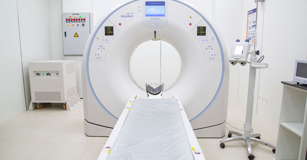
A researcher at KTH Royal Institute of Technology in Stockholm is developing sharper computed tomography (CT) imaging technology that could make radiotherapy more precise and personalised for cancer patients, according to an interview published by Karolinska Institutet.
Mats Persson, a senior lecturer and associate professor in the Department of Physics at KTH and a member of the Medical Imaging Physics research group, said the goal is to create images that clearly show tumours before, during and after treatment, so radiation is delivered to the right place at the right dose.
"Today, 72 per cent of all cancer patients in Sweden are alive ten years after diagnosis. Since most receive radiotherapy, better treatment can help more people survive," Persson told Karolinska Institutet.
Persson's work focuses on photon-counting computed tomography, a newer imaging technique that he believes will produce clearer and more useful medical images over the next decade. He said this technology could help distinguish between different types of cancer and allow clinicians to plan radiotherapy more precisely. Other methods he is exploring include phase-contrast imaging, which uses X-rays differently to produce higher-resolution images with less noise, and new technology for imaging radioactive tracers that can show how these substances move through the body over time.
The imaging technology has been developed through collaboration between researchers at Karolinska Institutet (KI) and KTH, specifically the teams of Staffan Holmin at KI and Mats Danielsson at KTH, Karolinska Institutet reported.
Persson is one of several KTH researchers contributing imaging expertise to the Centre for Medical Radiation Science, a collaborative initiative that brings together KTH, Stockholm University (SU), KI, and Region Stockholm. The centre aims to jointly develop future radiotherapy treatments and position Stockholm as a leading cluster in radiotherapy research.
"The radiotherapy of the future requires several different types of expertise," Persson said. "KTH develops the technology, Stockholm University contributes expertise in radiation physics, and KI and Karolinska University Hospital provide medical and clinical expertise. When technology developers, physicists and doctors work together, new solutions can benefit patients more quickly."
For students considering careers in medical physics, biomedical engineering, or radiation oncology, the centre represents a growing interdisciplinary field where technical and clinical expertise converge. The collaboration model means graduates with skills in physics, engineering, and data analysis could find opportunities in both research and clinical settings.
Persson said that over the next ten years, medical images will become both clearer and more useful. He envisions a future where information from medical images, tissue samples, and blood tests can be combined to choose the right combination of radiotherapy and medicines for each patient. Treatment could also be adjusted over time to maximise effect while minimising side effects.
"Patients may receive more personalised treatment," Persson said. "A world-class research and healthcare environment can also give patients in Sweden earlier access to new treatment methods."
The centre also provides industry partners with a natural collaborator and helps Swedish companies maintain their world-leading position in radiotherapy, according to Karolinska Institutet.